Hydronephrosis
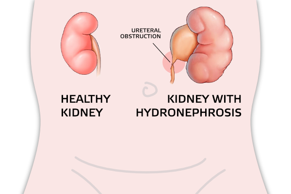
Hydronephrosis is a condition in which urine is retained in the collecting system of the kidneys, and is reflected in its expansion.
Renal pelvis (pyelonephritis) and renal calyces (calyx) are expanded. In case of great expansion, there is pressure on the kidney tissue (parenchyma). The most common cause of hydronephrosis is narrowing (stenosis) of the pyelouterine joint (the joint of the renal pelvis and ureter).
It develops prenatally while the kidney is still forming. Research confirms that it is more common in men and on the left side. However, both kidneys can be affected.
The narrowing prevents the normal flow of urine from the kidneys. This causes hydronephrosis (swelling of the kidneys). Prenatal ultrasound often diagnoses hydronephrosis as early as in the 15th week of fetal development. Early detection allows assessment and treatment soon after birth.
Signs and symptoms
Symptoms of UPJO may be abdominal mass (which is felt at a routine examination by a primary care physician), or a urinary tract infection with fever, abdominal or back pain. Back pain may be even more pronounced with increased fluid intake.
UPJO symptoms may not appear until the obstruction progresses. High-grade kidney obstruction can lead to kidney damage and loss of kidney function. Sometimes obstructions are found after an injury to the back or abdomen and the ultrasound image shows an enlarged kidney.

How is UPJ joint stenosis diagnosed?
The first step in diagnostics is anamnesis. Some of the diagnostic tests and examinations include:
- Ultrasound of the abdomen and urinary tract
- Magnetic urography
- Kidney scintigraphy/dynamic
- Urinary cystourethrography is performed in order to exclude reflux (return) of urine to the kidney
Treatment
Treating this condition requires corrective surgery called pyeloplasty. The purpose of treatment is to improve urine flow and prevent further damage to the renal parenchyma. Sometimes it is necessary to place a stent, catheter or percutaneous nephrostomy immediately in order to, at least temporarily, eliminate the symptoms of urinary incontinence.
The goal of pyeloplasty is to remove the narrowed part of the ureter. The operation is performed under general anesthesia. Towards the end of the procedure, an internal ureteral stent, also known as a "double J" stent, is placed. The stent is usually removed 3 weeks after surgery and the treatment is completed.
Return to normal physical activity is expected four weeks after surgery.
