alternative

Urology

A pediatric urologist provides diagnostic, medical, and surgical expertise for pediatric disorders affecting the urinary and genital tract.

The most common problems of children that fall under the domain of urology are genital abnormalities and bladder disfunction.

When to see a urologist?

Urologist’s consultations are necessary for new-borns if there was suspicion of the presence of kidney and urinary tract diseases during pregnancy. If your child’s genitals do not look or function properly (e.g. tight foreskin in boys), if there is swelling or pain, schedule an appointment.


What do we provide?

In addition to anamnesis and review of medical documentation, our urologist will perform an examination and give advice for further treatment and possible additional diagnostics (laboratory analysis, ultrasound, etc.), prescribe therapy or perform necessary procedures in order to eliminate the problem.


One of the most common problems faced by parents of boys is the problem of pulling the foreskin (prepucium) over the head (glans) of the penis. This condition is called phimosis, and can have two forms.

Primary phimosis (physiological) is a natural phenomenon, present in all boys at birth. The foreskin cannot be pulled over the glans at this time because it is attached to it by a series of small joints (adhesions), which break over time. Primary phimosis is a natural process that usually resolves on its own, or with the help of a doctor if its dynamic is slow.

Secondary or pathological phimosis occurs when the opening of the foreskin is too narrow and cannot be pulled over the head, or when scar tissue is created, due to trauma or violent pulling of foreskin. This problem requires treatment, application of creams and gradual pulling of the foreskin, and sometimes the only solution is circumcision.

It is very important that parents do not act on their own concerning this problem, because they can make the situation worse. It often happens that the parent pulls the foreskin by force thinking that it is a quick solution. Violent pulling of the foreskin can cause bleeding, infection, but also secondary phimosis or paraphimosis.

Paraphimosis is a condition in which the foreskin remains stuck below the edge of the glans and cannot be pulled back. This can be a serious problem because it leads to swelling of the glans and restricts blood flow. It requires an urgent visit to the doctor, and can even lead to surgery.

At Medikid, we solve the problem of adherent glans skin by adhesiolysis, under local anesthesia. Anesthetic cream is applied to the skin in order to make the separation of adhesions painless. After a successful intervention, proper hygiene and regular exercise are mandatory so that adhesions do not re-form, on which the parent receives detailed instructions and advice.

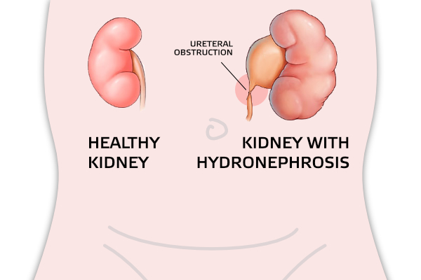
Hydronephrosis is a condition in which urine is retained in the collecting system of the kidneys, and is reflected in its expansion.

Renal pelvis (pyelonephritis) and renal calyces (calyx) are expanded. In case of great expansion, there is pressure on the kidney tissue (parenchyma). The most common cause of hydronephrosis is narrowing (stenosis) of the pyelouterine joint (the joint of the renal pelvis and ureter).

It develops prenatally while the kidney is still forming. Research confirms that it is more common in men and on the left side. However, both kidneys can be affected.

The narrowing prevents the normal flow of urine from the kidneys. This causes hydronephrosis (swelling of the kidneys). Prenatal ultrasound often diagnoses hydronephrosis as early as in the 15th week of fetal development. Early detection allows assessment and treatment soon after birth.


Signs and symptoms

Symptoms of UPJO may be abdominal mass (which is felt at a routine examination by a primary care physician), or a urinary tract infection with fever, abdominal or back pain. Back pain may be even more pronounced with increased fluid intake.

UPJO symptoms may not appear until the obstruction progresses. High-grade kidney obstruction can lead to kidney damage and loss of kidney function. Sometimes obstructions are found after an injury to the back or abdomen and the ultrasound image shows an enlarged kidney.


How is UPJ joint stenosis diagnosed?

The first step in diagnostics is anamnesis. Some of the diagnostic tests and examinations include:

  • Ultrasound of the abdomen and urinary tract
  • Magnetic urography
  • Kidney scintigraphy/dynamic
  • Urinary cystourethrography is performed in order to exclude reflux (return) of urine to the kidney

Treatment

Treating this condition requires corrective surgery called pyeloplasty. The purpose of treatment is to improve urine flow and prevent further damage to the renal parenchyma. Sometimes it is necessary to place a stent, catheter or percutaneous nephrostomy immediately in order to, at least temporarily, eliminate the symptoms of urinary incontinence.

The goal of pyeloplasty is to remove the narrowed part of the ureter. The operation is performed under general anesthesia. Towards the end of the procedure, an internal ureteral stent, also known as a "double J" stent, is placed. The stent is usually removed 3 weeks after surgery and the treatment is completed.

Return to normal physical activity is expected four weeks after surgery.

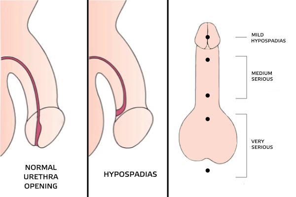
Hypospadias is a common structural difference of the penis where the urethra (the tube that carries urine from the bladder to the outside of the body) is located on the underside, not the tip of the penis. The opening can be located anywhere on the lower surface of the penis up to the scrotum.

A condition called chordee is often seen in hypospadias. Chordee is the descending curve of the penis. This can happen with or without hypospadias.

Types of hypospadias include:

  • mild (urethral opening is located on the head of the penis, no curvature of the penis is present - glandular, coronary)
  • moderate (urethral opening is located just below the head of the penis or in the middle of its body, there is a slight curvature of the penis - subcoronary, penile)
  • very heavy (urethral opening is located at the base of the genital organ, or on the scrotum, is accompanied by a large curvature of the penis - penoscrotal, perineal)


Hypospadias occurs in one in 150-300 boys. If a boy has hypospadias, there is a 15 percent chance that his brother will have the condition. Eight percent of fathers who have a son with hypospadias also have the condition.

The exact cause of hypospadias is unknown. There are many factors believed to be involved in its development. Genetics, the environment and hormones can be the reasons that contribute to the development of hypospadias.

Hypospadias is usually noticed at birth. Together with the incorrectly placed opening, the foreskin is often incomplete and uneven, ie. hypospadiform rearranged. It's called a dorsal hood.

Treatment of hypospadias is surgical.

In order to better understand vesicoureteral reflux, we will say a few words about the normal structure and function of the urinary tract.


Urinary tract

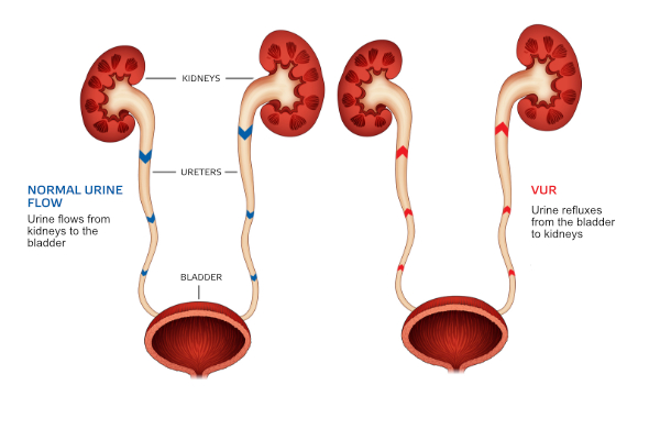
Kidneys clean and filter the blood to produce urine. Urine is a liquid waste. It travels from the kidneys through the ureters into the bladder. The bladder retains urine. It acts as a storage container. As the bladder fills, the muscle relaxes in order to enable the bladder to contain more urine. The control muscle (sphincter) remains firm to prevent urine leakage.

Inside the bladder, a valve mechanism prevents urine from returning to the ureters. When the bladder fills up, it sends a message to the brain. The brain decides when to start releasing urine. The bladder contracts as the control muscle relaxes. This allows the bladder to expel all the urine.


Reflux

What is VUR? Vesicoureteral reflux (VUR) is a condition in which urine can flow from the bladder back to the ureter and kidney. This is caused by a problem with the valve mechanism.


The pressure of urine filling the bladder should close the ureter. It must not allow urine to flow back into the ureter. When the ureter connects to the bladder at an unusual angle, reflux can occur. This can also happen when the length of the ureter that passes through the bladder wall is too short.

VUR becomes a problem when urine in the bladder becomes infected. Infected urine travels back to the kidneys. This can cause a kidney infection. Kidney infections cause damage.


How is VUR diagnosed?

VUR may be suspected if the ultrasound shows an enlargement of the ureter or renal pelvis. In order to diagnose VUR, the so-called urinary cystourethrography or MCUG in necessary. This is an X-ray scan of the urinary tract using a special color (contrast). If VUR is present, this test also shows its degree. There are five degrees of VUR.

Recently, VUR can also be diagnosed by contrast ultrasound cystography. A device is inserted into the bladder, which is visualized on a special ultrasound device. This method can determine the degree of VUR.


How VUR is treated and monitored...

...depends on the degree of the reflux itself, as well as the general condition of the child, their orderly growth and development.

It is possible to monitor VUR by introducing uroprophylaxis (antibiotics can be used to prevent infections until VUR recedes on its own).

Endoscopically - "the gold standard" in VUR treatment, performed by injecting endo paste at the place where the ureter enters the bladder. Children usually go home the same day.

Finally, surgically - the so-called ureteral re-implantation, which aims to correct ureteral defects and stop VUR.